
Respiration – Shortness of breath – Emergency management in Alpha-1
Author
Heike Isensee and co-author Dr. Alexander Rupp
Our lungs are a high-performance organ – in a healthy person, they process the equivalent of a hot air balloon full of air every day. Without breathing (and therefore without oxygen supply), irreversible damage occurs after just 3 minutes without oxygen.
Shortness of breath can have many causes:
- Infections (caused by viruses, bacteria, rarely fungi or mixed forms)
- Allergic reactions
- Environmental factors (e.g., particulate matter/smoke/soot) or lower oxygen concentrations, e.g., at an unusual altitude.
- Physical exertion
- Other additional illnesses (comorbidities), e.g. heart failure
- Smoking (the inhaled carbon monoxide occupies oxygen transporters and causes inflammation/emphysema)
- Acute or chronic inflammation of the airways
- Chronic destruction of the alveoli in the lungs (emphysema)
- Psychological stress, especially anxiety. Anxiety causes shortness of breath, shortness of breath causes anxiety! (Vicious cycle)
Peak flow, cough and mucus
How can I recognize a deterioration in my condition before acute shortness of breath occurs?
- Regular peak flow measurement: A decline in your usual peak flow reading could indicate an impending problem, even if you feel healthy. Use the peak flow meter while standing upright and holding the device horizontally. The reading indicates your maximum exhaled airflow in liters per minute. It is recommended to take measurements twice (up to three times) in the morning, at midday, and in the evening. More frequent measurements are advisable if you have a bronchial infection, are starting new medications, or if the readings fluctuate significantly. For patients with respiratory conditions, an escalation (increase) of inhaled therapy is usually already discussed with your doctor.
- Increased mucus production
- Thicker mucus than usual
- Increased coughing
Notes on this: Coughing is a natural reflex to mobilize mucus, which is produced, for example, due to smoking, inflammation, dust, etc., and transport it out of the airways with the help of cilia. Yellow-green phlegm suggests a bacterial infection (only then is antibiotics advisable, and only after consulting a doctor). Whitish phlegm is more indicative of a viral infection. The color of the secretions can be used as a clue, but should not be overemphasized as a diagnostic tool. A better way to differentiate is, for example, by measuring C-reactive protein (CRP) in the blood. Proper coughing technique is important. Never try to forcefully cough up mucus. The mucus should be breathed slowly and effortlessly up to the larynx and then expelled by clearing the throat. If possible, always cough with pursed lips, inhaling after every two to three coughs. Prolonged coughing fits without phlegm should be suppressed as much as possible.
Alpha-1 bronchitis carries a high risk of chronic bronchitis. This involves increased mucus production, and at the same time, the cilia may be damaged or destroyed, for example, in smokers or ex-smokers. As a result, mucus clearance becomes increasingly difficult.
Important: Every patient should be able to recognize a deterioration in their condition and manage it themselves. This includes increasing inhaled therapy as agreed upon with the doctor, using appropriate breathing techniques (pursed-lip breathing), and adopting breathing-easing postures. Patients should also be able to independently use their emergency inhaler if their condition worsens, increase their maintenance medication as prescribed, or, in cases of severe deterioration, take a course of oral corticosteroids (see also the action plan below).
Emphysema and pursed-lip breathing
In emphysema, the lung tissue is flaccid because the delicate walls of the alveoli are destroyed and the walls of the small airways (bronchioles) are hypermobile. The bronchi are also flaccid. A whistling sound during exhalation ("stridor") indicates obstruction of the lower airways (because the bronchi collapse, preventing proper airflow, similar to deflating a balloon while pinching the inlet). This leads to progressive overinflation of the lungs, as more and more air becomes trapped. By creating counter-pressure, for example, through pursed-lip breathing during exhalation, the alveoli can be emptied more effectively, as the bronchi remain open.
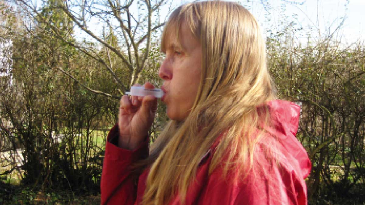
The correct exhalation pressure for pursed-lip breathing can be practiced as follows: Remove the (often green) plunger, i.e., the part used to push, from a 10ml disposable syringe. Then, close your lips around the wider, rear end of the syringe and exhale through it.
Correct, controlled exhalation using pursed-lip breathing leads to a nearly complete exhalation. This allows for better inhalation.
Tips for better mucus clearance:
- Drink plenty of fluids (note: for example, if you have kidney disease, you should not exceed the amount you drink; please follow the instructions of your doctor).
- Light pats on the back
- Take a deep breath
- Exercise (endurance training and strength training)
- Expectorants (especially natural ones, e.g., containing thyme, ivy, or primrose) can be helpful. In some individuals, pharmacological expectorants may lead to increased mucus production; in such cases, they should not be used.
- Respiratory physiotherapy
- Vibration massage, e.g. with a vibration device or manually by respiratory physiotherapists
In case of deterioration (unstable lungs or infection):
- Drug therapy, usually with cortisone in tablet form for 5 days; in case of bacterial infection, antibiotics may also be prescribed in consultation with the doctor.
- Nebulizers with, for example, saline solution (to make yourself: boil 9g of salt with 1 liter of water, or 1 level (not heaped!) teaspoon with 0.5 liters of water, which produces an isotonic saline solution) or in ampoule form as a pre-prepared solution. In cases of constricted airways, possibly combine with short-acting beta-agonists or anticholinergics (e.g., salbutamol or ipratropium bromide as a ready-to-use inhaler).
- For acute mucus buildup, use aids such as the Flutter or RC Cornet. (These devices should not be used for prevention, but only for treatment, i.e., when mucus is actually present). A good effect can also be achieved by blowing bubbles through a straw in a glass of water.
Exacerbation
An exacerbation is defined as an acute worsening of symptoms requiring a change in therapy. This occurs frequently in COPD stages II-IV, especially in winter (due to infections, air pollution, but also due to unknown causes).
There are 7 warning symptoms of an acute exacerbation:
- Signs of an infection (fever, sore throat, runny nose, fatigue, etc.)
- Increased coughing in general
- Nighttime coughing or shortness of breath that keeps you awake
- Reduced resilience
- Increased shortness of breath (usually first noticed during physical exertion)
- Reduced peak flow value
- Emergency spray needs to be used more frequently than normal.
Note: The rescue inhaler should be used early; therefore, the term "emergency inhaler" is misleading. An immediate improvement in breathing should be noticeable after inhaling the spray; otherwise, another problem exists.
Change in therapy during exacerbation:
- Increase in bronchodilator therapy (sprays) or additional inhalation via the jet nebulizer, so.
- Anti-inflammatory drug therapy, i.e., cortisone, usually in tablet form
- In case of purulent sputum, blood test results indicating inflammation, fever, or changes on the chest x-ray (infiltrate): antibiotics
- In case of severe shortness of breath: oxygen (via mask / non-invasive ventilation)
- In case of poor general condition, very severe shortness of breath, increasing respiratory insufficiency (i.e., the oxygen level in the blood is much too low, the CO2 level much too high): hospital admission.
Symptoms of severe shortness of breath
These could include, for example:
- The breathing is rapid and shallow; the patient is anxious or panicky.
- Tightness in the chest, possibly pain
- Audible breathing sounds
- The skin is pale or blue-violet, especially on the lips and fingernails.
- The patient tries to support himself with his arms in order to instinctively activate the accessory respiratory muscles.
- Often cold sweat
Other acute lung emergencies
Pneumothorax
- Symptoms: acute pain in the side of the chest, radiating into the shoulder
- Acute, persistent shortness of breath
- A gurgling sound in the chest; the pain subsides over time.
- Weak to no breathing sounds on that side
What may have happened: The negative pressure in the narrow space between the outer lining of the lung (pleura) and the inner lining of the chest wall (pleura) is disrupted by an influx of air, for example, from ruptured emphysematous bullae. This causes the lung to collapse, as it lacks a stable shape. Usually, a tube (drain) must be inserted into the pleural space for a few days to re-expand the lung.
Pneumonia
- Symptoms: slow, breathing-dependent increase in pain
- Fever, general feeling of illness
What may have happened: Germs (bacteria, viruses, rarely fungi) have entered the lungs and are causing inflammation with increased fluid production.
embolism
- The pulse rate is higher than 100/min at rest, and blood pressure is frequently low (systolic < 100 mmHg).
- The respiratory rate is massively increased, shortness of breath
- Bloody sputum, pain when breathing in
- Life-threatening condition
What may have happened: a blood clot (thrombus) blocks a pulmonary artery. The thrombus may have traveled to the lungs from completely different parts of the body. The resulting failure of the affected lung area leads to acute oxygen deficiency.
Action plan
The action plan describes the medications that must be taken at different stages of health.
A distinction is made between the stable lung phase, in which the "usual" medications are needed, the unstable lung phase, in which the dosage of medications may need to be increased, and the infection phase, in which further medications, e.g. cortisone in tablet form or antibiotics, become necessary.
The treatment plan must be drawn up by the treating physician. It is important to have the necessary medications on hand. These should always be taken along on vacation, etc. Every patient should be able to recognize when they are transitioning from a stable to an unstable phase and then adjust their therapy accordingly, as instructed by the doctor.
Emergency plan:
It should also be prepared by the doctor and always carried by the patient.
The following information is important:
- The patient has Alpha-1
- What medications does the patient receive in emergency situations? (Name and dosage)
- Indication of potentially other serious illnesses
- Contact details of relatives and the doctor
- Reference to any existing advance healthcare directive
- Information on the peak flow values that are normally measured
You should also always carry your emergency inhaler! There is a European emergency card available as a template in various languages. This can be obtained (usually for a fee) from pharmacies, many general practitioners, or online. It is advisable to carry such a card when traveling abroad.
Better breathing: how can I breathe more easily?
- Pursed-lip exhalation during exertion
- Breathing-easing posture
- Take a deep breath before strenuous activity
- Exhale during exertion, inhale during relaxation.
The breathing-easing postures serve to additionally activate the accessory breathing muscles, which hardly have to do any work during "normal" breathing.
These are various muscles in the shoulders, neck, and back. Because the body no longer has to support the weight of the arms, more energy is available for these muscles.
Breathing-relieving positions – along with pursed-lip breathing and emergency inhaler – are the best prerequisite for getting better airflow in case of acute shortness of breath.
Positions that ease breathing
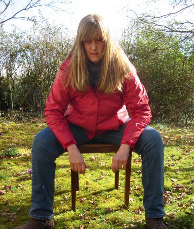
Coachman's seat: Sit on a chair with your legs hip-width apart, optionally with one corner of the seat tilted forward. Lean your upper body slightly forward with a straight back, supporting yourself with your hands or elbows (reaching almost to your knees).
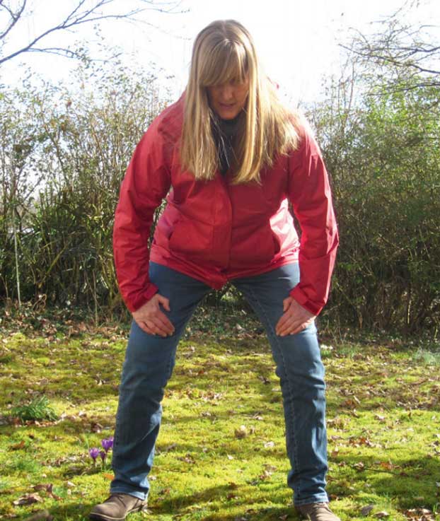
The goalkeeper position: Squat slightly, upper body leaning slightly forward, back straight. Again, the upper body is supported with the hands on the legs (slightly above the knees).
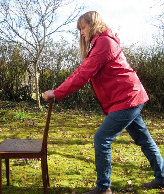
Support while standing For example, using both hands against a chair back (or walking sticks): In a staggered stance, the upper body is slightly inclined forward with a straight back, and the arms support the upper body against the chair back, etc. Support can also be achieved with just one hand to the side.

„Student seat“ (only works on a chair with a backrest): Sit „backwards“ on the chair, rest your forearms on the backrest and your head on your arms.
Tasks of the first aider
- Calm the patient; panic should be avoided at all costs.
- Position the patient sitting and still (the patient should not move).
- Tight clothing, especially around the neck, opens
- Be careful when "fresh air" by opening a window: especially for asthmatics, a sudden influx of cold air can trigger or worsen bronchospasm. For allergy sufferers during pollen season, large amounts of pollen entering the room can exacerbate their condition.
- Regularly check the patient's level of consciousness by speaking to them or touching them.
- Make bystanders clearly aware of the emergency situation
- If the situation does not improve immediately: Don't hesitate, call an ambulance.
- Clearly assign tasks: Who calls 112, who goes outside the building to guide the ambulance service to the patient, who stays there in pairs for possible resuscitation?
- When calling emergency services at 112, calmly and completely provide the following information: Who/Where/Alpha-1 patient/acute respiratory distress (or respiratory arrest) and wait for follow-up questions from the emergency dispatch center (do not call on your own).
hang up!) - Check your breathing regularly
- If no breathing is detected, begin resuscitation measures (30 chest compressions in the center of the chest, followed by 2 rescue breaths, repeat until emergency medical services arrive. The rhythm of the Bee Gees' song "Staying Alive" is considered ideal for this). You can't really go wrong with resuscitation. The biggest mistake is not performing resuscitation when there is no circulation.
It is important that the first responder remains calm and collected. Before an emergency even occurs, the person affected should inform their family members, colleagues, and friends about what to do in an emergency.


