
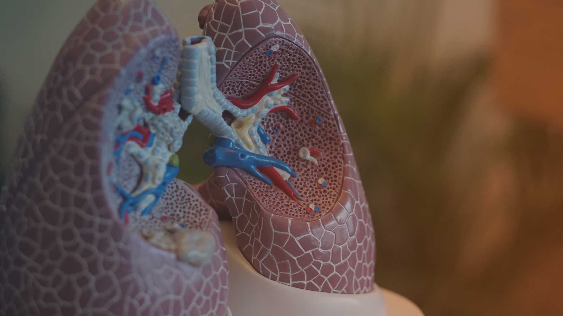
Lung volume reduction
Author
Speaker: PD Dr. Daniela Gompelmann, Thorax Clinic at Heidelberg University Hospital
Summary: Prof. Dr. med. Gratiana Steinkamp, as published in Alpha1 Journal 1-2019.
COPD disease
The two main components of COPD are the narrowing (obstruction) of the airways and the lung overinflation caused by emphysema. Furthermore, the lung disease also has significant effects on the entire body outside the lungs and, for example, leads to weight loss.
During pulmonary function testing, the reduced airflow can be represented, among other things, by the FEV1 (forced expiratory volume in one second) measurement. Every person with AATD should know their FEV1 value. In cases of obstruction, this value is usually reduced, i.e., below 801 Tp3T of the predicted value. Only in the early stages of a mild obstruction is this value still considered normal. Lung hyperinflation can be estimated using the residual volume (RV). The RV measures the amount of air that remains in the lungs after maximal exhalation. Values above the normal range are pathological. While the normal range is not precisely defined, a measurement above 130 or 150 Tp3T of the predicted value is generally considered indicative of lung hyperinflation.
Lung hyperinflation and its consequences
People with pulmonary hyperinflation and the resulting enlarged rib cage have altered breathing mechanics. This means they have to exert significantly more muscular effort for normal breathing, even at rest, compared to healthy individuals, which creates a feeling of shortness of breath. In particular, the enlarged, air-filled lungs push the diaphragm, the most important respiratory muscle, downwards towards the abdominal cavity. The downward-pressed diaphragm flattens its shape and therefore moves less efficiently.
Reduction of lung volume
Severely overinflated lung areas do not contribute sufficiently to breathing and gas exchange. Instead, they compress adjacent, otherwise functioning lung areas and also alter respiratory mechanics. As early as the 1950s, thoracic surgeons began removing severely emphysematous lung areas to allow the healthier lung areas to expand more effectively. While the surgical results at that time were encouraging, they were associated with high mortality, leading to the procedure being largely forgotten.
It wasn't until the 1990s that lung volume reduction surgery became more common again. Study results from this period demonstrated not only an improvement in quality of life but also longer survival for the operated patients. However, mortality after this procedure remained high, averaging 81 Tp3T within the first three months post-surgery (Fishman et al. NEJM 2003). Two very small studies on AATM were also published, which likewise showed an improvement in lung function and exercise tolerance, but were also associated with a trend toward increased mortality compared to conservative therapy (Cassina et al. Eur Respir J 1998; Stoller et al. Ann Thorac Surg 2007).
Based on these results, alternative treatment options were sought that could achieve the same positive effect as lung volume reduction surgery, but with lower mortality.
Valves for lung volume reduction
The idea was to reduce the amount of air in a lung lobe without the need for surgery. Various techniques were developed that can be performed non-surgically during a bronchoscopy. One such method involves the implantation of valves, which are placed in the most emphysematous and overinflated lung lobe. Due to their one-way function, these valves prevent new air from entering the lobe during inhalation. However, during exhalation, air flows out of the lung through the valves. Over time, the air content in this lung lobe decreases, and the tissue gradually collapses (a condition known as atelectasis). Consequently, adjacent, healthier lung sections can expand again, and the diaphragm can move more freely.
In each patient, doctors first use a computed tomography (CT) scan to determine which of the five lung lobes is most overinflated and damaged by emphysema. During a bronchoscopy, the valves are then inserted into the bronchi leading to the affected lung lobe. This procedure takes about 20 minutes.
Study results from approximately 1,000 patients have now been published in randomized clinical trials (e.g., Klooster et al. NEJM 2015; Criner et al. Am J Respir Crit Care Med 2018). When patients are carefully selected, their FEV1 increases and residual volume decreases after valve implantation. Furthermore, physical performance, such as the 6-minute walk test, and quality of life improve. Based on these promising results, experts have also recommended lung volume reduction with valves in treatment guidelines, most recently the German COPD guideline of 2018.
Indication for valves
Valve therapy can only be considered in patients with confirmed emphysema on CT scan. For lung function testing, specific threshold values must be met, such as FEV1 between 15 and 501 TP3T of predicted and RV above 2001 TP3T of predicted. Guidelines also apply to walking distance and quality of life. It is crucial that patients have otherwise received optimal conservative treatment. Furthermore, valve therapy is only promising if there is no collateral ventilation between the lung lobes. Such collateral ventilation occurs when adjacent lung lobes are not completely separated but are connected by a tissue bridge. In such cases, the lobe treated with the valve could be ventilated from the neighboring lobe, thus negating the valve's effectiveness. Physicians can detect collateral ventilation using CT scans or a special measurement during bronchoscopy (Chartis®).
However, there are other reasons that argue against valve therapy. For example, the patient must not have bronchiectasis, chronic lung infections, acute pneumonia, or any suspicious findings in the lungs that require further investigation.
Treatment results after valve therapy
Carefully selected patients can benefit from valve implantation: their exercise tolerance, quality of life, and lung function improve, and symptoms decrease. Life expectancy is also extended if maximum volume reduction of the target lung lobe is achieved (Gompelmann et al. Respiration 2018). However, the lung does not recover completely after the procedure, and the progression of COPD cannot be stopped.
An important and relatively common complication occurs in about one-third of patients within the first three days: a pneumothorax. The lung tissue of the adjacent lobe tears due to the shift in volume, allowing inhaled air to flow through the tear into the chest cavity. The resulting loss of negative pressure in the chest cavity causes the lung to collapse. This leads to severe shortness of breath and intense pain. Sometimes, air is even forced out of the chest cavity and can be felt under the skin as a crackling bulge.
A pneumothorax is treated by inserting a drain from the outside into the chest cavity between two ribs, allowing the escaped air to be suctioned out. This works well in most cases. However, if not enough air can be drained, the drains must be removed. In this case, the patient gains no benefit from the drain therapy. If the drains do not need to be removed and the pneumothorax heals with drainage alone, the chances of success are good even after this complication.
In two small studies, valve therapy was also used in a total of 21 individuals with AATD (Tuohy et al. Clin Respir J 2012; Hillerdal et al. Respiration 2014). The lung function value FEV1 increased significantly and rapidly after valve therapy and remained at a better level than before the procedure for years in most patients.
Summary
In selected patients with COPD and pulmonary emphysema, valve therapy can significantly improve quality of life. Experience with AATD in patients is still very limited. The potential advantages and disadvantages of the valve must be carefully weighed by the physician and patient. It is important to note that valve therapy does not preclude lung transplantation, which should be considered, especially in younger patients with AATD and already significantly impaired lung function.
Summary: Prof. Dr. med. Gratiana Steinkamp, as published in Alpha1 Journal 1-2019.


